Cardiac Ultrasound (Echocardiography)
Ultrasound is a non-invasive, modern technique that produces a visual imprint of the interior of the body. It allows the doctor to achieve a depth of detail that is not possible with X-rays.
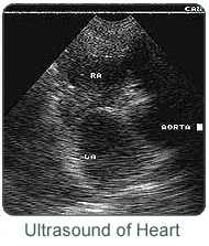
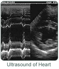
Echocardiography is a standard, non-invasive technique for assessing cardiac anatomy, pathology and function. High-frequency sound waves produced from a hand-held device are directed at the heart. The sound waves are reflected back to the machine and an image is produced. This image is transmitted to a video monitor where it can be seen. The image may be still or moving.

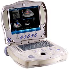
Portable Veterinary Ultrasound Machine
Ultrasound uses sound waves, is non-invasive, painless, uses no dies and has no obvious harmful effects. It is perfect for diagnosing many types of heart problems.

The heart is a very complex organ. It has four valves, an electrical conduction system, and four chambers, all of which are crucial to its function. As in people, animals suffer from a wide variety of heart diseases. In order to correctly diagnose and treat these diseases, it is necessary to see inside the heart, visualize the movement of the chambers, and to look at the valves in motion. This is the area where ultrasound predominates. Using ultrasound, it is possible to watch the heart in motion and to measure each of its' parts. Ultrasound also allows the doctor or technician to measure the actual physical effects of various heart medications and to carefully adjust dosages.

Most heart diseases are treatable, and in most cases, animals can live normal lives once the nature of their condition is under control.
[ Search Articles ] [ Article Index ] [ Previous Page ]